
‘Doing a PhD is never boring!’ How Guido Stam built a microscope that can measure bacteria without causing harm
A microscope with incredible sharpness that leaves samples unharmed – Guido Stam helped develop one. During his PhD research, he combined light and electrons to study biological samples. ‘We can now measure things that simply weren’t possible before.’
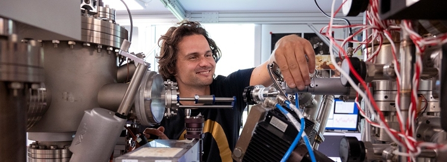
What Guido Stam enjoys most about doing a PhD in experimental physics is the variety of the work. ‘It’s not just thinking all the time – you also have to get hands-on with the microscope. And our microscope produces stunning images that I get to analyse for scientific data. I really enjoy that part.’
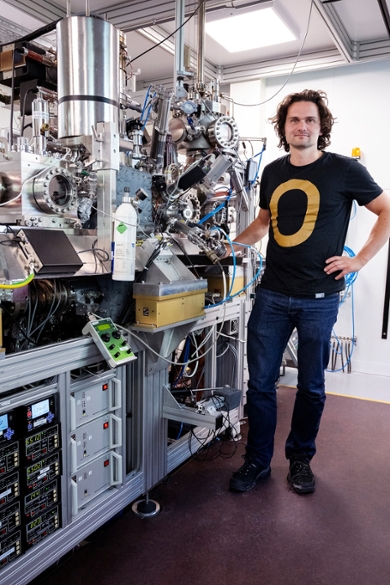
Over the past four years, Stam contributed to developing a new kind of microscope: Optical Near-field Electron Microscopy (ONEM). It brings together the best of both worlds – the high resolution of electron microscopy, and the ability to examine samples without damaging them, like you can with a traditional light microscope. The research was funded by an EU Horizon 2020 Grant.
Applications in biology and electrochemistry
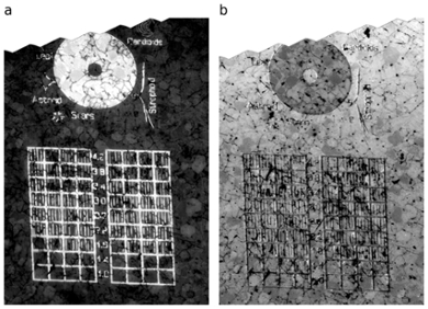
To prove the concept works, Stam and his colleagues placed E. coli bacteria – known for causing intestinal infections – under the microscope and took measurements. Afterwards, they checked the same sample using an optical microscope and saw the bacteria were still very much alive.
Stam says: ‘Our microscope also works well in liquids, which is great for electrochemical experiments like those in the ANION programme, coordinated by the Leiden Institute of Chemistry. A regular light microscope doesn’t zoom in far enough, and an electron microscope has the resolution but its high-energy electrons can trigger unwanted chemical reactions that interfere with the experiment. Our microscope works completely differently and avoids those issues. It’s based on an idea by Austrian physicist Thomas Juffmann, which we developed further together and his PhD students.’
‘The resolution of our microscope is already seven times better than that of a conventional light microscope. Our goal is to reach twenty times better and measure accurately down to ten nanometres.’
Turning light into an electron stream
Stam explains how the microscope works: ‘We shine light onto the sample from behind. As the light passes through the sample, it gets altered slightly. That light then hits a thin photocathode, which converts it into a stream of electrons. Where less light hits the photocathode – because something like a bacterium was in the way – fewer electrons are released. These electrons are then accelerated and turned into an image using the electron optics of our low-energy electron microscope (LEEM).’
Persistence paid off
Designing and getting the photocathode to work was the biggest challenge of Stam’s PhD. ‘For three years, I regularly hit roadblocks – failed experiments, technical problems. But when I finally produced a really clear image, I was over the moon. Everything came together in the final months, and we managed to carry out a whole series of successful experiments.’
Summary of the research results:
- A measurement with a resolution of 31 nanometres
- First successful electrochemical experiment
- A successful measurement of E. coli bacteria, with no damage to the cells
A delicate balance
The ONEM microscope is producing better results all the time. Stam says: ‘It’s a very delicate system. The sample needs to be perfectly aligned in the microscope – too much or too little light can ruin the measurement. Then the photocathode has to be applied just right, and finally the whole setup must be stable enough for the electron optics to work properly. Every day, we're learning more about all the variables we need to control.’
Passing the torch
Stam wrapped up his research at the Leiden Institute of Physics with the defence of his PhD thesis, held at the Academy Building on 4 June 2025. The project continues, and a new PhD student has already started working on further improving the ONEM technique for electrochemical experiments.
The differences between microscopes
Microscopy is the science of visualising matter on a scale too small to see with the naked eye. But every type of microscope has its technical limits.
The light microscope is the most familiar, which can clearly image objects down to around 200 nanometres.
For smaller structures, we turn to the electron microscope. A high-quality electron microscope can achieve a resolution of 0.05 nanometres – sharp enough to distinguish individual atoms. The downside, however, is that it uses high-energy electrons for imaging, which can damage delicate samples like bacteria or proteins.
Stam is helping to establish a new technique: Optical Near-field Electron Microscopy (ONEM). ‘The resolution of our microscope is already seven times better than that of a conventional light microscope, and it doesn’t harm biological samples,’ he explains. The aim is to improve this further – ideally reaching twenty times the resolution of a light microscope, and the ability to measure with an accuracy of ten nanometres.