

In pictures: animal mummies in a scanner
The story of Tutankhamun, the Egyptian pharaoh, is world famous. But did you know that the Ancient Egyptians mummified not only people but animals too? The National Museum of Antiquities in Leiden recently put a bunch of animal mummies through a CT scanner. This was in collaboration with Canon Netherlands and researchers from Leiden University.
Mention mummies and you’re likely to think of majestic pharaohs given an elaborate royal burial. All embalmed and well, their body would be safely stored in a rock tomb in the Valley of the Kings, while their spirit set off on its journey through the afterlife. The good preservation methods would enable them to stay there for eternity – or at least until an archaeologist or grave robber discovered their burial place.
But what not everybody knows is that the Ancient Egyptians also mummified animals on a large scale. This was occasionally the favourite pet of a rich man or woman, but was usually an animal sacrifice. These mummified animals were sold as votives in temples that visitors could offer to the gods. This was a way to keep the gods happy, which was handy if you wanted to get a good job or have a healthy child, for instance.
Animal mummies as revenue model
‘In those days a whole industry had sprung up around this custom,’ says Lara Weiss, curator of the Egyptian collection at the National Museum of Antiquities (RMO). ‘You have to imagine a continuous supply of mummified animals being sold in the temples. These animals were bred and killed with the sole aim of mummification. It was a real business.’
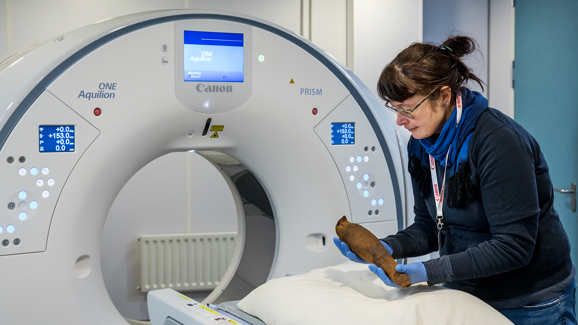
RMO has an impressive 73 animal mummies in its collection, from crocodiles to cats and falcons to fish. When some of these mummies were going to travel to Japan as part of a touring exhibition, the museum decided to make 3D scans of their insides. Weiss: ‘This meant that the Leiden public would still be able to see the mummies. And it also gave us the opportunity to look at high resolution at the insides of the mummies, and to see if we could discover anything new about them.’
Snake mummy in the scanner
Weiss’s first port of call was Berend Stoel. He is a professor of image processing and knows everything about the ‘techie side’ of the CT scanner, as he puts it. His regular work consists of writing programs that automatically analyse or even interpret images from scanners. He looks for arthritis, lung disease and eye disorders, for instance.
Outside of the LUMC’s regular opening times, he is willing to place something rather different in the CT scanner. He once scanned none other than a Stradivarius to see if the annual rings in the famous violin’s wood are responsible for its superior sound. (They aren’t). And for Naturalis Biodiversity Center, he recently scanned a dinosaur bone.
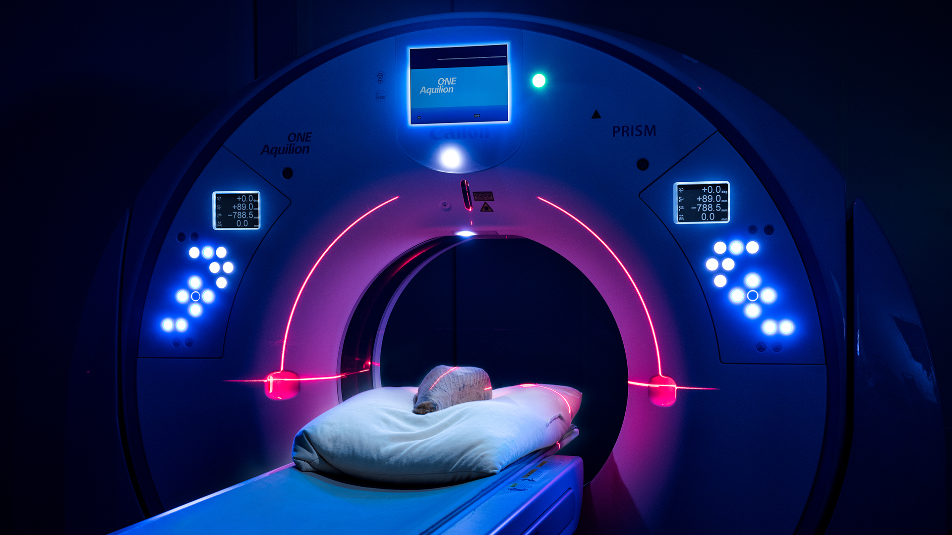
He was therefore unfazed when asked in December 2019 to scan a snake mummy. The assignment: make a 3D image of the snake in the highest possible resolution. No mean feat, apparently. A snake’s spine consists of minuscule structures, which make it incredibly difficult to find the right settings to bring these into focus.
Dummy fish head
In the end Stoel’s colleague Irene Hernandez-Giron came up with an inventive solution. She bought a fish from the fishmonger, boiled it and made a model to practise with. Stoel used this ‘dummy mummy’, wrapped up in a sock, to get the CT scanner’s settings ready for the real work. The fragile snake mummy from the RMO could then be scanned straight away and returned to the museum pronto.
Weiss then called on two Leiden biologists, Michael Richardson and Merijn de Bakker. They specialise in the developmental biology of vertebrates, or animals with a backbone. They were given the noble assignment of working out from the CT scan what kind of snake it was. That still wasn’t clear because you can’t just unwrap a mummy. Perhaps they would be able to see this from the shape of the fangs. This can give away the species, especially in combination with geographical information about where the snake was found.
The researchers were less fortunate here. The fangs were not quite visible enough to determine the snake’s species. Richardson’s intern and snake expert Roel Wouters suspects that it is an Egyptian cobra, however. He can tell this from the characteristic shape of the skull and the snake’s prevalence in Egypt.
-
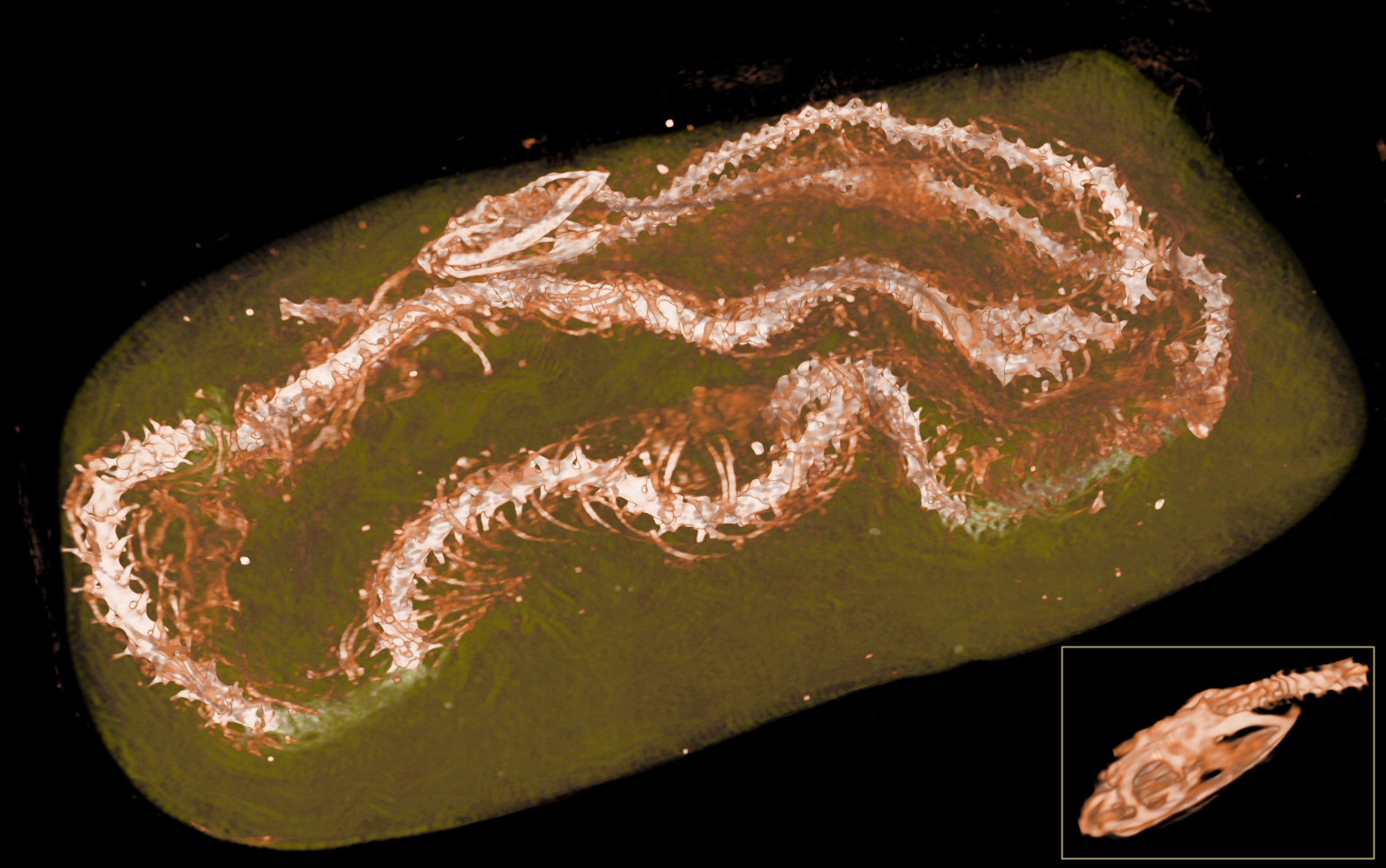
Scan of a snake mummy -
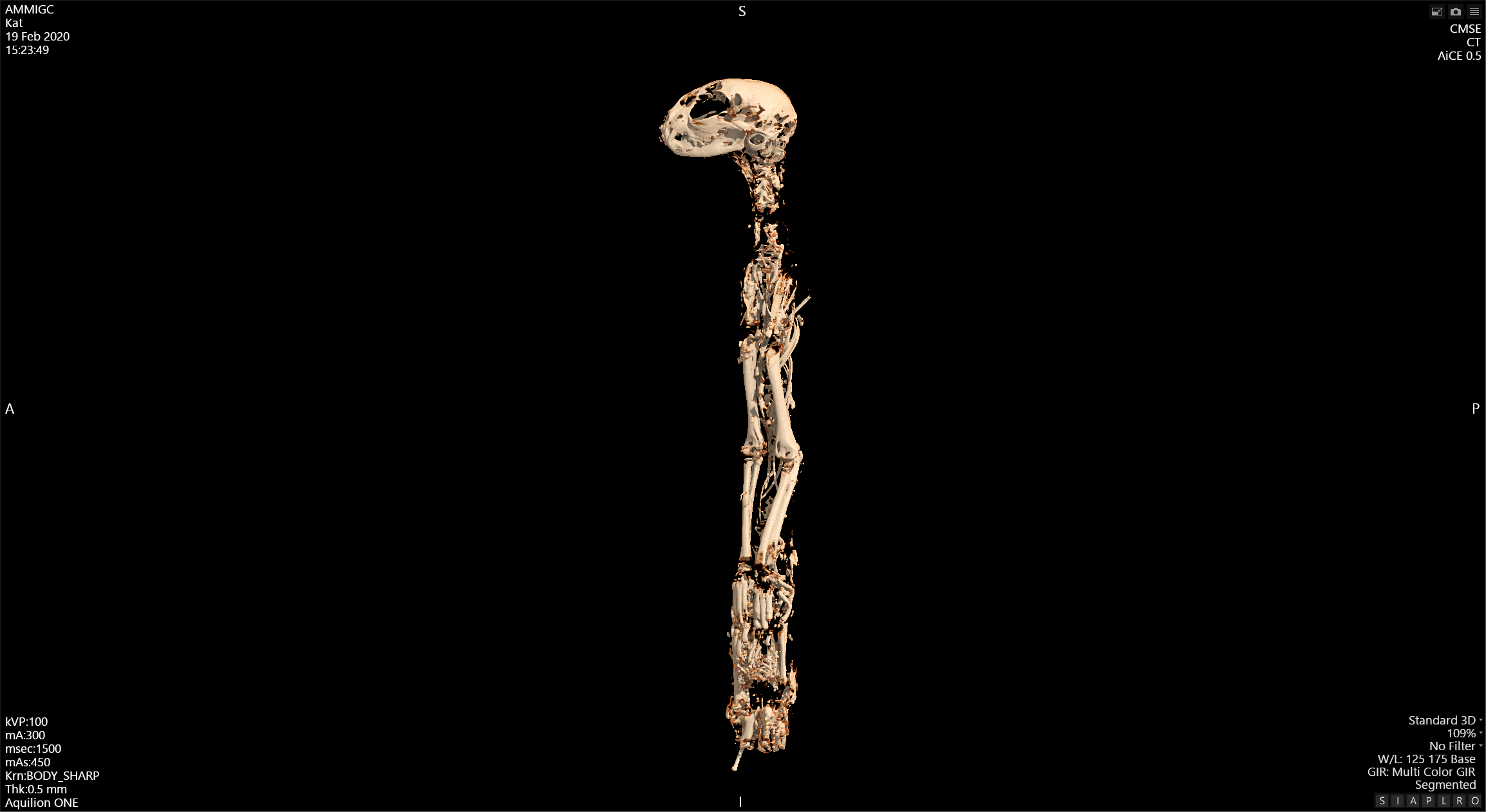
A cat -
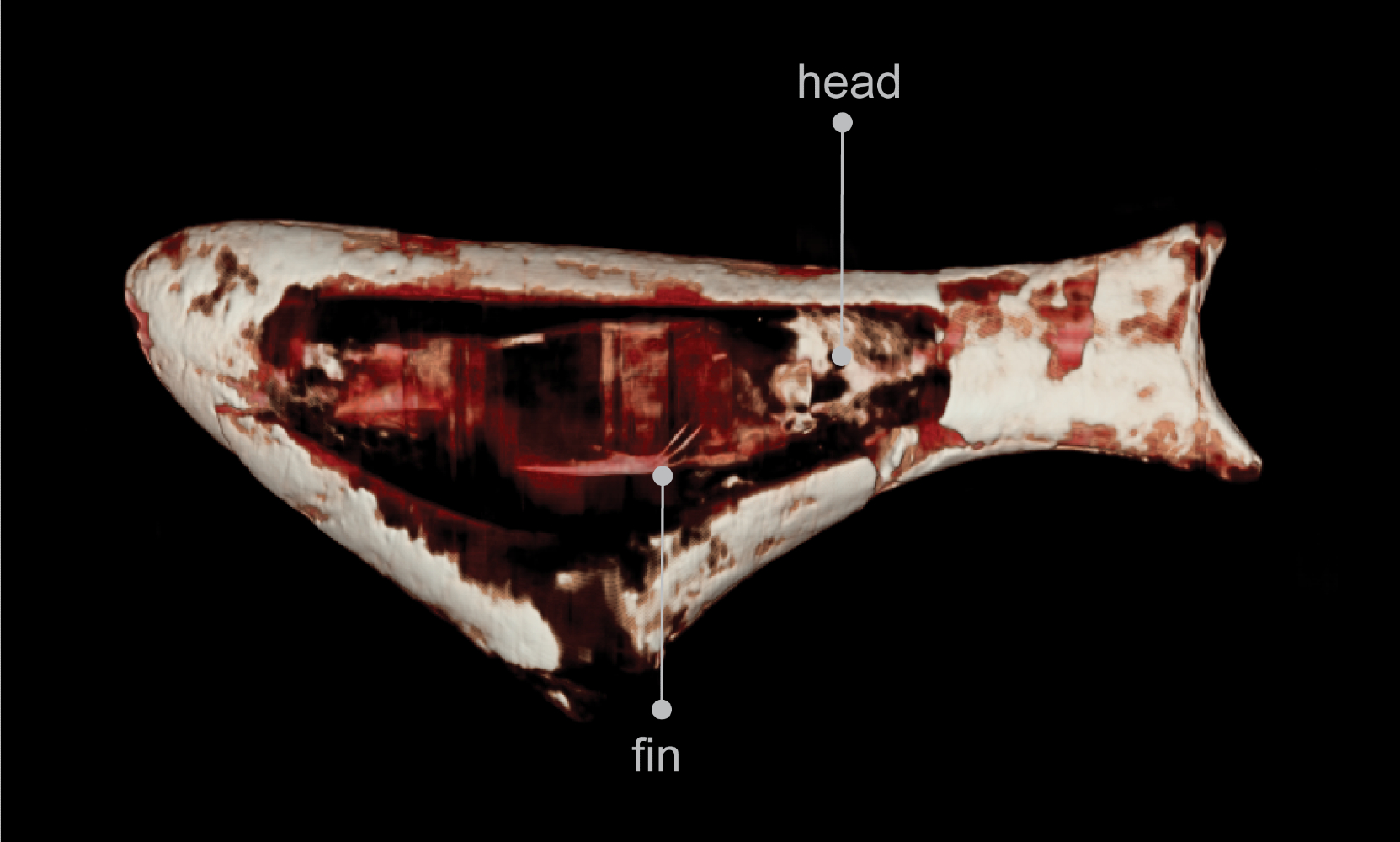
A fish -
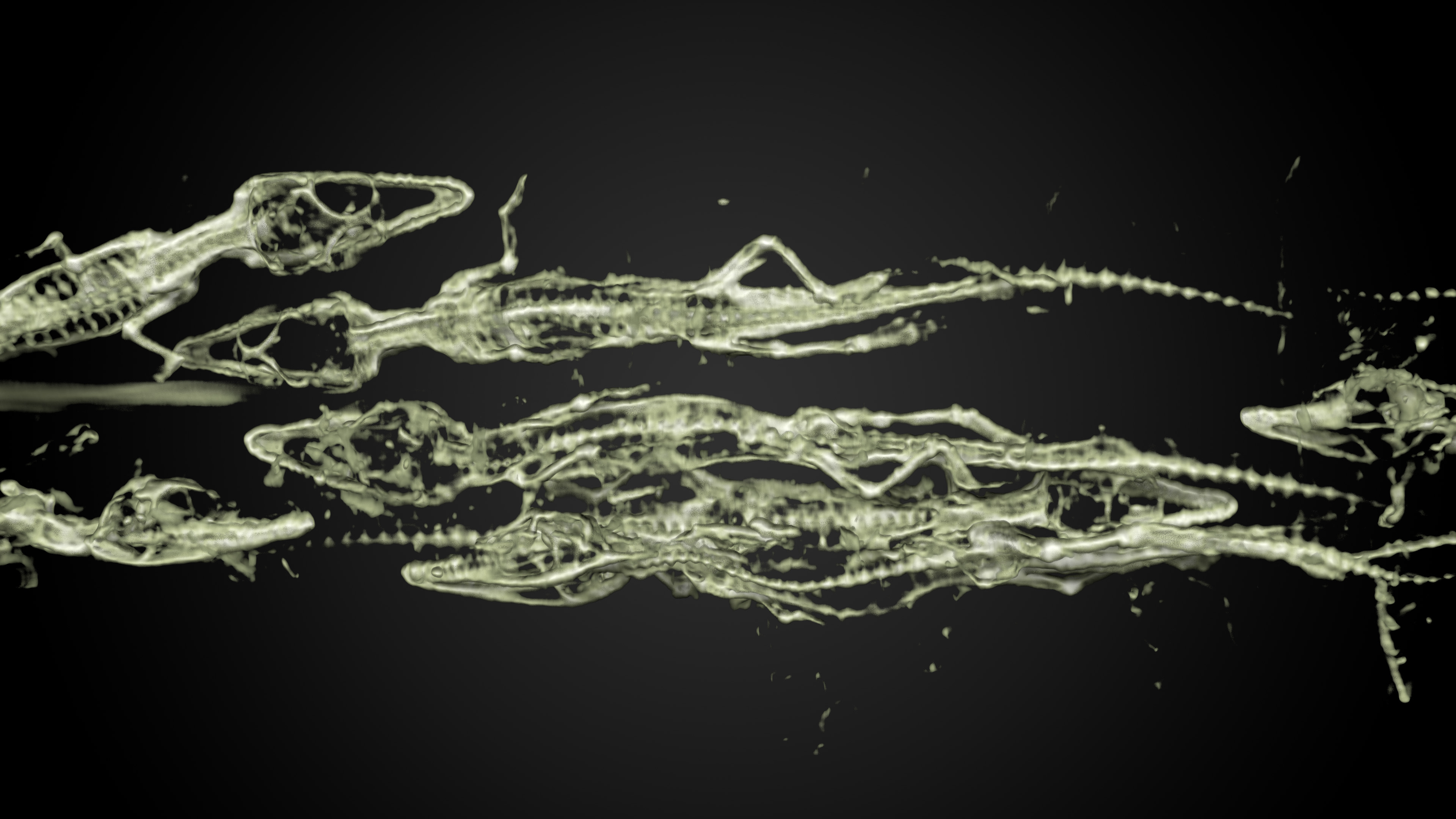
Baby crocodiles -
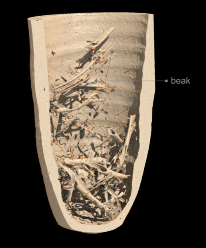
Ibis mummy inside a pot
Hours puzzling
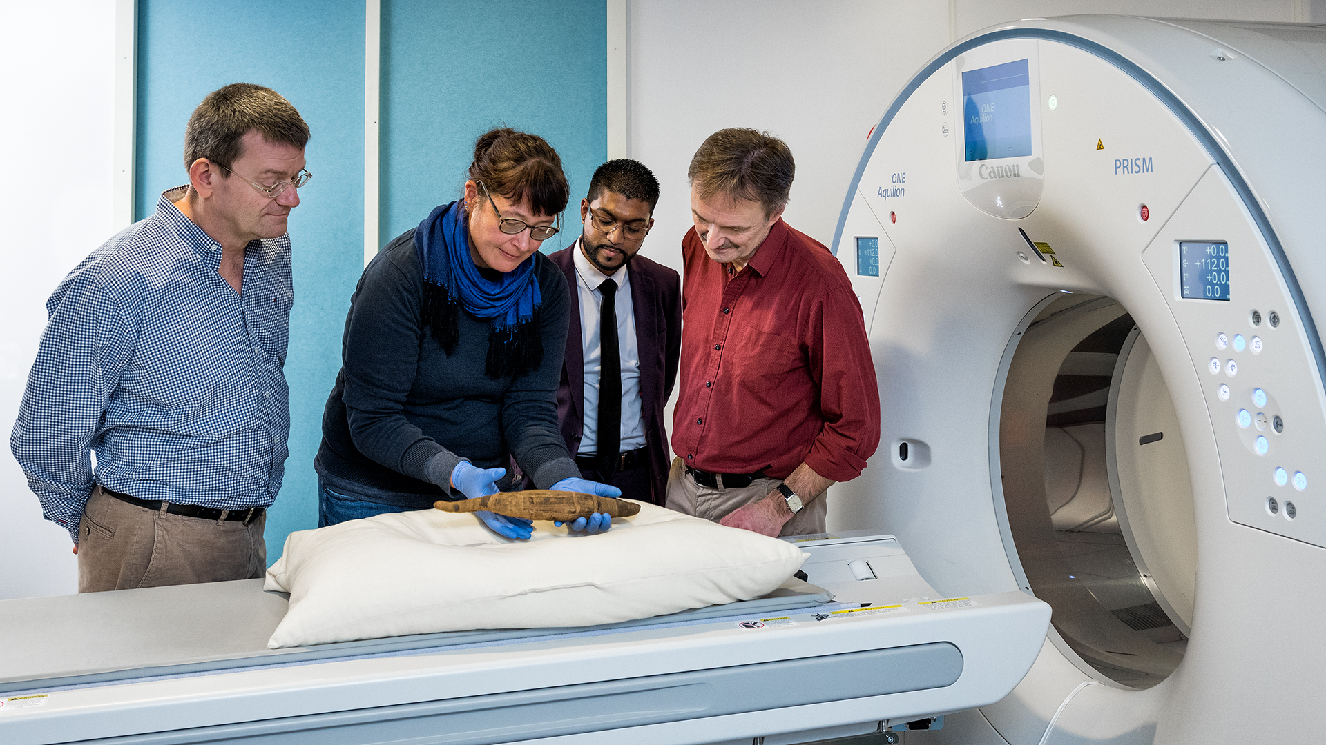
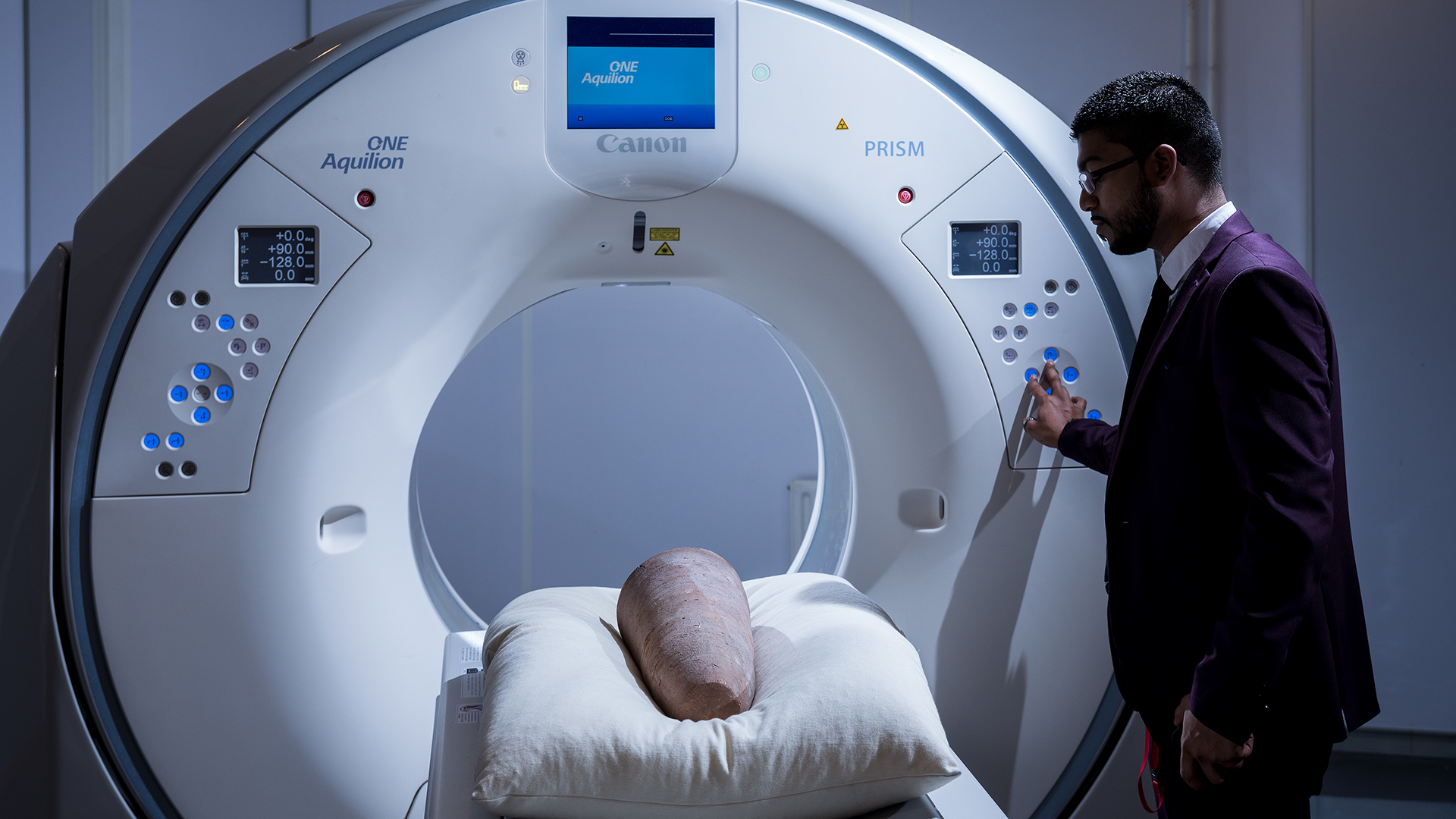
In the months that followed, this team effort between archaeologists, CT experts and biologists was repeated several times. After the snake mummy had been fed into a scanner, so too were crocodile, dog, fish, falcon, ibis and cat mummies. As the project needed completing quickly because of the Japanese exhibition, these animals were not scanned in the LUMC but in a demonstration scanner in Zoetermeer belonging to Canon. This company builds medical equipment and saw scanning animal mummies as a good way to demonstrate the functionality of its latest CT scanner. This produced beautiful scans of the ancient mummies.
For Richardson and De Bakker, the scans resulted in hours of puzzling. Many of the animals proved to have been shaken around over the centuries. The falcon’s bones were oddly folded, and its head was missing even. The ibis skeletons also proved incomplete and to have undergone somewhat of a battering. Nonetheless, the biologists could see from the bent beak that it was indeed an ibis.
The cat mummy was in a better state, but here the biologists made a rather sinister discovery. The animal had been young and healthy when it was offered to the gods. Before the poor animal was mummified, its neck had been wrung; the position of the neck vertebrae was testament to this. Its body was then mercilessly flattened and wrapped.
Useful exercise
Both biologists are keen to emphasise that the mummy project was a more of a hobby, a fun distraction from their real work. Richardson: ‘In our everyday lives we study the motion system of snakes, for instance, so their muscles and vertebrae. Obviously it’s much easier to have a fresh snake rather than an ancient, mummified specimen that you’re not allowed to unwrap.’
‘But it’s a fantastic project to get a glimpse of what your neighbours are up to,’ Merijn de Bakker adds. ‘It’s the perfect example of an interfaculty project, and therefore gives you a good idea of what’s going on in other parts of the university.’
For Stoel scanning the animal mummies was also a fun jaunt. ‘I’ve earned a bit of a reputation for this,’ he smiles. ‘But patient care always takes priority, of course, which is why we do this work in the evening or at the weekend.’ Future LUMC patients may actually benefit from the escapades in the CT scanner: according to Stoel scanning dinosaurs or mummies is a good exercise. ‘It gives me new insights and keeps me on my toes.’
Text: Merijn van Nuland
Photos: RMO and Canon